Microscopy & Microtechniques
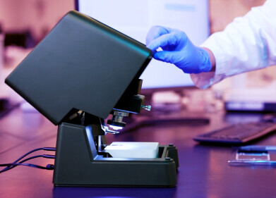
Fluorescence Imaging Microscope Released
Oct 01 2010
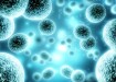
UVP, LLC exhibited at World Molecular Imaging Conference in Kyoto, Japan, to showcase the new iBox® Explorer™ Fluorescence Imaging Microscope along with UVP’s extensive line of iBox in vivo imaging systems for rapid high resolution analysis of fluorescently tagged cells, tissues, organs and whole animals in the visible and NIR wavelengths. UVP now offers three systems for real time, fluorescence imaging to speed up basic and preclinical research studies in tumours, oncology, heart and metabolic diseases.
The iBox Explorer is unique in its ability to micro image cells and organs subcutaneously and within the body cavity of living mice. The iBox Explorer joins the whole mouse imagers iBox Scienta and iBox Spectra in revolutionising in vivo fluorescent imaging. The iBox Explorer operates through its intuitive software control using optical configurations that are parcentered and parfocal, allowing seamless imaging through the magnification ranges. Included is a leading-edge high frame rate cooled color camera with technology that enables quick detection, image capture and high throughput. Applications of the iBox Explorer include imaging whole organs, tumor/host margins and interactions, tumour microenvironment, vasculature, and micro metastases.
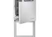
UVP’s line of BioImaging Systems includes the BioSpectrum® multifunctional imaging systems which combines a high resolution CCD camera and advanced features for enhanced imaging fluorescent and chemiluminescent imaging capabilities.
Digital Edition
ILM 49.5 July
July 2024
Chromatography Articles - Understanding PFAS: Analysis and Implications Mass Spectrometry & Spectroscopy Articles - MS detection of Alzheimer’s blood-based biomarkers LIMS - Essent...
View all digital editions


24_06.jpg)


Events
Jul 30 2024 Jakarta, Indonesia
Jul 31 2024 Chengdu, China
ACS National Meeting - Fall 2024
Aug 18 2024 Denver, CO, USA
Aug 25 2024 Copenhagen, Denmark
Aug 28 2024 Phnom Penh, Cambodia