-
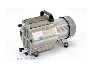
 Fig. 1: BioSpectrum Imaging System
 -
 Fig. 2: Chemiluminescent blots of rabbit sera dilution series comparing film to BioSpectrum CCD capture and results (inset CCD) are more sensitive than film.
 -
 Fig. 3: CCD and film exposure by a calibrated light source, GLO21, to compare sensitivity.
Laboratory products
Digital Chemiluminescent Imaging of Protein Blots
Aug 04 2010
No Film Required for Chemiluminescent Imaging
BioSpectrum® Imaging System (Fig. 1) is ideal for imaging chemiluminescent protein blots. Protein blotting is a routine technique for determining the presence or absence proteins and can provide additional information on the antigen amount in the sample. The use of chemiluminescent imaging of protein blotting and the sensitivity of this procedure compared to film is discussed here. The results show that the BioSpectrum with the cooled BioChemi 500 camera and an f/1.2 lens are superior to film in sensitivity, accuracy, dynamic range, speed, and simplicity. Due to the high sensitivity and resolution of the BioSpectrum, the resultant images are both quantitative and publication quality.
Immunoblotting reactions using chemiluminescent substrates are captured with higher sensitivity using the BioSpectrum with cooled CCD camera as compared to film (Figure 2). This is confirmed using quantitative low light standards (Figure 3). Weak signals are poorly recorded or missed completely with film, while the BioSpectrum camera effectively records weak and strong signals of the blot and standard (Figures 2 and 3).
Because the BioSpectrum acts as a benchtop darkroom, there is no need for a full traditional darkroom. Once positioned on the imaging platform, the membrane is focused, door closed and image captured with a one button preset. Images were processed with VisionWorks® LS image acquisition and analysis software to remove background signal and quantitate the intensity of the bands. Rapid preview simplifies setting exposure times, and the acquired image is available for immediate and accurate quantitation. The chemiluminescent exposure typically takes from 10s to a few minutes. Northern and Southern blots generally require exposures ranging from 5 to 30 minutes. Unlike fluorescent blotting applications, no excitation light is needed.
In contrast, film requires a full darkroom with running water and has many more steps: while in the full darkroom, the film needs to be removed from light tight packaging and placed in film cassette. The membrane, in plastic wrap, is placed on top of the film and gentle pressure applied, usually by closing the film cassette, to ensure good contact with the film. Any movement of luminescent blot while situating on the film will result in spurious contact exposure creating a double image on the film. Once processed, the film must be scanned to quantitate the bands on the blot. Due to the very limited dynamic range of film, film is easily saturated by bright signals and at the same time will not record weak signals, giving poor data accuracy (figures 2 and 3).
Chemiluminescent imaging with the BioSpectrum is simple, fast and straightforward, yielding full 16 bit images for quantitation and publication. Chemiluminescent blots offer very high sensitivity, rivaling radiolabeled techniques, and can be easily reprobed without worrying about radioactivity disposal. Rapid, high-resolution image capture is possible through the use of cooled CCD camera and f/1.2 low light lenses.
Digital Edition
Lab Asia 31.6 Dec 2024
December 2024
Chromatography Articles - Sustainable chromatography: Embracing software for greener methods Mass Spectrometry & Spectroscopy Articles - Solving industry challenges for phosphorus containi...
View all digital editions
Events
Jan 22 2025 Tokyo, Japan
Jan 22 2025 Birmingham, UK
Jan 25 2025 San Diego, CA, USA
Jan 27 2025 Dubai, UAE
Jan 29 2025 Tokyo, Japan
.jpg)


