Laboratory Products
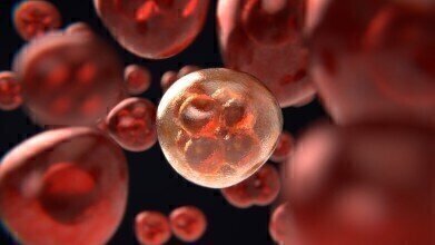
How Do Cancer Cells Spread?
Oct 03 2020
Breakthrough research from Johns Hopkins Medicine has revealed new insight into how cancer cells spread, with the findings published in the journal Cancer Research. The process is called metastasis and sees malignant cells infiltrate blood vessels to spread from a primary tumour to other parts of the body. In a major leap forward for cancer research, the team used tissue engineering technology to replicate a three-dimensional blood vessel and simulate the metastasis process. After artificially growing breast cancer cells nearby, the team observed cancerous cells overtaking part of the blood vessel and releasing a cluster of malignant cells into the bloodstream.
“We observed that cancer cells can rapidly reshape, destroy or integrate into existing blood vessels,” explains senior study author f the study Andrew Ewald, Ph.D. “Just as people going scuba diving versus ice climbing require different tools, cancer cells bring different equipment depending on the job they intend to perform," says Ewald, a professor of cell biology at the Johns Hopkins University School of Medicine and co-director of the Cancer Invasion and Metastasis Program at the Johns Hopkins Kimmel Cancer Center. “Determining what that equipment is can help us understand how to stop cancer.”
Mapping tumour microenvironments
Initially, Ewald and the team expected to observe clusters of up to 10 cells released migrating from a tumour and attaching to blood vessel walls via protein barriers. Instead, they saw parts of existing tumours commandeer blood vessel walls, then release cancer cells directly into the bloodstream.
“We never saw that," says Ewald. "What we kept seeing instead was that a piece of an existing tumour would take over a neighbouring blood vessel wall, putting cancer cells in direct contact with the circulation, and that the cancer cells could do so in a matter of hours. They didn't have to invade past the blood vessels; they became the blood vessels, and could just release cancer cells there."
Understanding “mosaic” vessels
The team refer to the newly infiltrated vessels as “mosaic” vessels, a reference to the mix of both natural and cancerous cells. As well as being present in around 6% of human breast tumours, these “mosaic” vessels are also found in melanoma skin cancers, gastric cancers and brain tumours known as glioblastomas.
Moving forward, Ewald says the three-dimensional model could be used to develop a deeper understanding of tumour microenvironments and how cancers spread.
Want to know more about the latest scientific breakthroughs? ‘UV-Vis: 5 steps to Pharmacopoeia compliance’ spotlights one of the most widely used instrumental techniques in pharmaceutical analysis.


Digital Edition
ILM 49.5 July
July 2024
Chromatography Articles - Understanding PFAS: Analysis and Implications Mass Spectrometry & Spectroscopy Articles - MS detection of Alzheimer’s blood-based biomarkers LIMS - Essent...
View all digital editions

24_06.jpg)


Events
ACS National Meeting - Fall 2024
Aug 18 2024 Denver, CO, USA
Aug 25 2024 Copenhagen, Denmark
Aug 28 2024 Phnom Penh, Cambodia
Sep 04 2024 Chiba, Tokyo, Japan
Sep 04 2024 University of Warwick, Coventry, UK