-
 Dr Fernando Perez-Cota
News & Views
Using Sound Waves to View inside Cells
Oct 14 2018
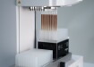
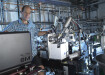
Dr. Fernando Perez-Cota, Faculty of Engineering at the University of Nottingham has won a prestigious five-year fellowship to explore the use of harmless sound waves to view deep inside living cells to aid early diagnose in diseases such as cancer. He is building a unique imaging instrument that uses sub-optical-wavelength sound (or phonons), typically used in the semiconductor and consumer electronics industries; however their use in scientific imaging is something new.
Dr Perez, of the Optics and Photonics Group, explains: “Many existing optical imaging techniques fail because they disturb or kill cells in the imaging process, especially with the use of toxic chemical dyes. Sound, by comparison, is harmless to life. Ultrasound, for instance, is the only safe method to image living human embryos.
“To exploit this at a cellular level, phonons are the right choice as their wavelength are in the nanometre range. This use of microscopic ultrasound is currently unexploited in the life sciences and healthcare, but it has many potential benefits in 3D imaging, which is something I will be investigating on the project.”
The new ultrasound microscope will use short laser pulses to generate phonons which spread into cells. The signal picks up and feeds back variations in ‘hardness’ (elasticity) of cell features (membrane, nucleus, etc) to build up a more detailed 3D picture that would be perceptible using light.
The phonon tool will allow scientists to observe the mechanical properties inside living cells in much higher resolution than with current optical or acoustic methods - resulting in better picture quality - and over more sustained periods of time. Researchers can also use the tool to study how abnormal and healthy cells may respond differently to drugs, changes in temperature, diet and atmosphere.
While the phonon microscope will support greater understanding of many issues in cell biology, Dr Perez intends to focus his research on health problems such as cancer, parasitology, osteoporosis and fertility. “There is a great deal of evidence that the elasticity of cancer cells differs significantly to normal cells, however capturing the traits of these cells in microscopic detail has proved challenging in the past. The phonon tool could potentially aid earlier disease diagnosis or improve scientific understanding of how cells behave, mutate and spread in the human body,” Dr Perez adds.


.jpg)
Digital Edition
LMUK 49.7 Nov 2024
November 2024
News - Research & Events News - News & Views Articles - They’re burning the labs... Spotlight Features - Incubators, Freezers & Cooling Equipment - Pumps, Valves & Liquid Hand...
View all digital editions


Events
Nov 18 2024 Shanghai, China
Nov 20 2024 Karachi, Pakistan
Nov 27 2024 Istanbul, Turkey
Jan 22 2025 Tokyo, Japan
Jan 22 2025 Birmingham, UK