-
 Harvard's latest microscopy method offers as-it-happens views of circulation
Microscopy & Microtechniques
Latest microscopy method offers video-rate cell visualisation
Dec 06 2010
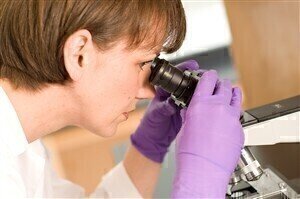
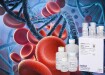
The latest microscopy method to be reported by Harvard University offers a real-time insight into the inner workings of cells and the human circulatory system.
Using stimulated Raman scattering, the latest microscopy technique to be developed by scientists at the academic institution is a means by which blood cells can be pictured passing through capillaries.
At present, surgery to remove lesions and tumours incurs a delay of around 20 minutes while excised samples are sent away for histological processing.
However, with real-time imaging, the Harvard experts believe this delay could be removed, or substantially shortened.
X Sunney Xie, one of the leaders of the project, says: "When we started this project 11 years ago, we never imagined we'd have an amazing result like this."
Harvard University says that its spectrum of learning experiences encompasses a broad range of both social and academic disciplines, with a "dedication to the pursuit of excellence" guiding its activities.
.jpg)
Digital Edition
Lab Asia 31.6 Dec 2024
December 2024
Chromatography Articles - Sustainable chromatography: Embracing software for greener methods Mass Spectrometry & Spectroscopy Articles - Solving industry challenges for phosphorus containi...
View all digital editions


Events
Jan 22 2025 Tokyo, Japan
Jan 22 2025 Birmingham, UK
Jan 25 2025 San Diego, CA, USA
Jan 27 2025 Dubai, UAE
Jan 29 2025 Tokyo, Japan